Two state universities collaborate to improve human and animal eyesight
02-25-09
Contacts:
Sinisa Grozdanic, Veterinary Clinical Sciences, (515) 294- 4900, sgrozdan@iastate.edu
Randy Kardon, University of Iowa and Veterans Administration Hospitals, (319) 356-2260, randy-kardon@uiowa.edu
Tom Ligouri, College of Veterinary Medicine, (515) 294-4257, ligouri@iastate.edu
Dan Kuester, News Service, (515) 294-0704, kuester@iastate.edu
Two state universities collaborate to improve human and animal eyesight
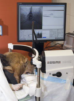
New imaging equipment scans the eye of an animal patient. What is learned in the Iowa State University College of Veterinary Medicine is being used by human doctors in Iowa City.
AMES, Iowa -- Iowa State University's College of Veterinary Medicine recently installed the newest generation of retinal imaging equipment for examining the eyes of animal patients.
Dr. Sinisa Grozdanic, an assistant professor of veterinary clinical sciences at Iowa State's College of Veterinary Medicine, is conducting research with Dr. Randy Kardon, a doctor and professor at the Veterans Administration and University of Iowa Hospitals and Clinics in Iowa City.
"What we're doing and what we've been doing in the last seven years is basically translational medicine," said Grozdanic. "We try to combine and apply knowledge from human medicine and veterinary medicine for the benefit of human patients and veterinary patients. There are so many diseases that are present in veterinary patients which are almost identical to human diseases."
The new imaging instrument is called spectral domain optical coherence tomography and is similar to ultrasound. Instead of sound waves, however, it uses coherent light waves, which give cross-sectional images of the layers of the living eye with much greater detail.
Kardon hopes the results of the research will help his human patients and Grozdanic's animal patients as well.
"Dr. Grozdanic and I are studying mechanisms of damage and new treatment of eye disorders that occur in both animals and humans," said Kardon. "The new structural analysis we get from the scanner will help us bridge the gap between the animal studies and the human studies that we are conducting simultaneously."
Kardon says the Veterans Administration, which funded the scanner, hopes to advance new research and treatment of disorders that affect the retina and optic nerve in soldiers and veterans.
This is the best available equipment -- not just the best for animal eyes, but among the best for any eyes, animal or human -- according to Grozdanic.
The images produced by the machine are almost as good as the quality doctors can get using a microscope -- while not having to extract tissue from the patient.
The imaging machine captures images of the retina which is only 200 microns thick -- about one-tenth the thickness of plastic kitchen wrap.
Some problems the instrument will help diagnosis are glaucoma and different forms of degenerative and traumatic optic nerve diseases.
"We made a huge step forward in terms of quality of image and diagnostics that we can provide," said Grozdanic.
"This will be a big advantage over what veterinary medicine can offer human medicine," Grozdanic said.
Humans and animals have many of the same eye diseases, according to Grozdanic. Studying these diseases in animals gets faster results because of shorter animal life spans.
"There are so many interesting things we can find in animals," he said. "And the life span of animals is shorter, so these diseases develop much faster in animal than in human patients. This can allow us to evaluate efficacy of new treatments and bring them much faster to human patients suffering from different blinding diseases."
-30-